 | Neuronal Substructure
Price: 0
Description:
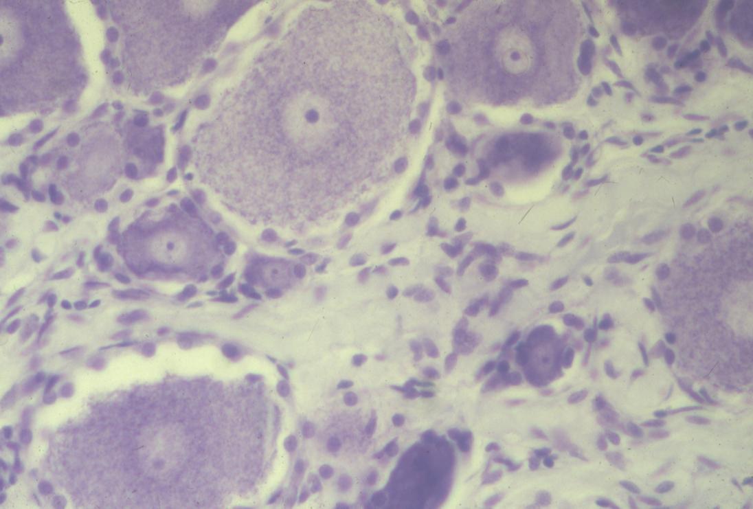
Light and electron microscopy are used to visualize intracellular structures of neurons and glia. Chemical staining reveals the combination of structural and chemical organization that compose neuronal and glial substructure. Nissl staining shows the location of the DNA and RNA structures in neuronal somata. Dorsal root ganglion (DRG) cell somata inter-twined with supporting and glial cells. Each DRG is evident by the light staining of the nucleus and the dark nucleolus (dot). The perineuronal supporting cells (satellite cells) ring each DRG soma. Elongated nuclei are of the myelin forming Schwann cells and mark the region where axons are passing into nerves. Note the variability in size of the DRG somata. These size differences are related to the diameter of axonal projections. Axons adjust the speed of conduction that is required for timing coincidence of signal arrival to each center."

|
